A new study led by chemistry researchers at the Case Western Reserve University has identified a method to detect inflammation in the body using antibodies, which could potentially lead to new blood tests for a range of conditions and diseases. The research, published in the Proceedings of the National Academy of Sciences (PNAS), has the potential to change the way inflammatory diseases like heart disease, Alzheimer’s disease, and certain cancers are diagnosed, and may also provide new insights for drug discovery.
“This research opens up an amazing number of pathways for future studies,” said Greg Tochtrop, PhD, a professor of chemistry at Case Western Reserve and the senior author of the study. “It will lead directly to better understanding inflammation and detecting diseases, as well as to discovering new drugs.”
In their research, Tochtrop and the team found that certain compounds formed from their interaction with reactive oxygen species (ROS) in a unique way allowing for detection by antibodies. ROS are highly reactive oxygen-containing chemicals that can damage DNA, proteins, and lipids. During inflammation immune cells produce ROS to kill bacteria and pathogens, but they can also be created by exposure to certain environmental conditions such as exposure to ultraviolet light, pollution, radiation, and smoking.
For this study, the team wanted to discover how ROS could react with the fatty acid found in all cell membranes called linoleic acid, forming compounds that can bind to RNA, DNA and proteins, called epoxyketooctadecenoic acids (EKODEs).
The investigators showed that the EKODEs react with nucleic acid cystine in a way that had never been described before and formed a stable bond. The compounds then accumulated in body tissues suffering from oxidative stress such as the brain, heart, liver, and other organs. Tochtrop then developed antibodies to these compounds from mouse models that were able to detect buildup of EKODEs in a variety of tissue from both mouse models and humans.
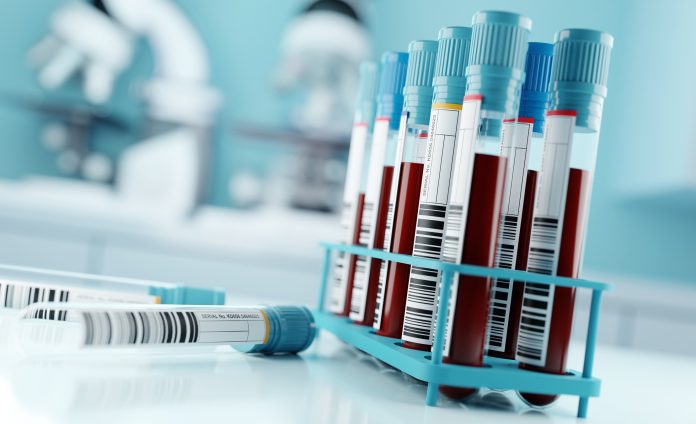
“What makes this so interesting and so potentially valuable is that we could detect unique compounds and concentrations in different tissues and organs, which means that you could potentially detect a variety of diseases with a blood test,” Tochtrop said.
The method could lead to blood tests similar to the A1C test for diabetes, which measures the percentage of hemoglobin coated with glucose over time. Just as the A1C test provides information on long-term glucose levels, an EKODE test could reveal oxidative stress levels in organs which would be indicative of disease progression.
The study’s findings also have implications for drug discovery. Since drug developers are often focused on identifying reactive cysteines to target in treatments, this research offers new insight into those cysteines. “Identifying reactive cysteines is central to drug discovery right now,” Tochtrop said. “This could help uncover many reactive cysteines that could be targeted for drug discovery, which is a valuable offshoot of our research.”
As this research progresses, the development of EKODE-based blood tests could become a new tool in clinical settings, allowing for more precise monitoring of inflammation and disease progression. The next phase of the work will involve refining the identification of EKODEs in different tissues and correlating these markers with specific disease states, further advancing the potential for both diagnostic applications and drug development.


