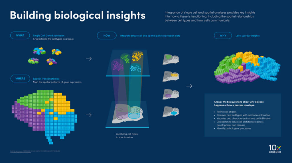
Biologists of all sorts seek the answer to this general question: What happens and where? The complexity of the answer and even the ability to address the question depends intimately on the problem being studied. A chain of technological advances over centuries, however, continues to give biologists tools to explore the what and where in a wide range of applications, from basic biology through healthcare.

chief medical officer, Mission Bio
Born as an observational science, biology started more with the where than the what of nature. “Understanding the spatial orientation of complex, heterogeneous human tissues has been of great interest since the advent of the first microscope over 500 years ago,” said Todd Druley, MD, PhD, chief medical officer at Mission Bio. “During that time, the ability to discern finer details of cells, their structures, and how they organize to perform a cumulative function has continuously improved.”
Along the way, scientists also learned how to analyze a sample’s genetic material. That started with bulk sequencing—looking at the total DNA in a sample containing many thousands of cells. Eventually, genomic tools made it possible to sequence a single cell’s genetic material, DNA or, more commonly, RNA. To answer what happens and where, though, scientists needed to combine single-cell sequencing and spatial technologies.

senior segment manager
cancer research, Illumina
“Spatial information has always been critical to research because a cell’s location within a complex tissue significantly influences its biology and the biology of the cells surrounding it,” said Robert Yamulla, PhD, senior segment manager, cancer research, Illumina. “Single-cell sequencing methods excel at maximizing information about individual cells, but unfortunately also involve dissociating those cells from their location within tissue.”
So, scientists developed ways to analyze a single cell’s molecular profile—from DNA and RNA to proteins—all while locking in its location in a sample. As Yamulla put it: “Combining spatial profiling with single cell allows researchers to understand both the spatial context of cells within tissue, while also allowing comprehensive measurement of multiple layers of regulation within individual cells.”
Making use of the combo

senior product marketing manager
10x Genomics
Such combinations of tools can be applied in many ways. Jyoti Sheldon, senior product marketing manager at 10x Genomics, provides a list of key applications: disease pathology and progression; developmental biology; neuroscience; immunology; drug discovery and development; and tissue engineering and regenerative medicine. For example, a tool that combines sequencing with spatial information “enables the detailed study of diseases, such as cancer, by identifying tumor heterogeneity, cancer stem cells, and the tumor microenvironment,” Sheldon explained. “Researchers can track how cells evolve and interact with their environment, aiding in the discovery of novel therapeutic targets and biomarkers.”
As another example, Druley noted: “Combining spatial transcriptomics or single-cell transcriptomics with single-cell DNA and protein expression could be an extremely powerful discovery tool to follow how changes in DNA alter RNA expression, subsequently impacting protein expression and how that disrupts healthy tissue structure.” The results might be used to develop new forms of precision therapy. “Although diagnostic tools that measure disease-specific DNA changes and/or protein expression have traditionally led the way into clinical care,” Druley said, “current tools can simultaneously measure point mutations, insertions-deletions, copy-number variants, and surface protein expression from thousands of cells.”

head of pathology, Nucleai
Scientists can also combine a range of datasets, such as proteomics data, to gather even more information. “While single-cell genomics gives us the full diversity of cells and cell states, proteins are the ultimate effectors of cellular function and a patient’s phenotype is directly linked with clinical outcomes,” said Kenneth Bloom, MD, head of pathology at Nucleai. “For example, the majority of the drugs being developed for cancer or immune disorders target proteins, and mapping proteins with spatial context offers a way to translate genomic findings into clinically relevant assays.” By combining all this information, scientists can zoom in on more specific aspects of biological mechanisms and where they take place, which could reveal extremely specific therapeutic targets.
Tweaking the technology
Although both single-cell sequencing and spatial analysis provide crucial information, scientists must work within the capabilities of the technologies. In brief, the sequencing can only analyze all of the genes within a sample or a specific set.

“One of the most important ways that cells interact is through physical proximity so spatial context is often important for solid tissues, however, many of the current spatial workflows rely on the use of targeted panels limited to a couple hundred genes whereas most single-cell approaches leverage either an unbiased approach with amplification based on the poly A tail of mRNAs or very large probe pools that target nearly the entire transcriptome,” said Steven Hoffman, senior staff, regional segment marketing manager, Illumina. “By integrating both single-cell and spatial preps together into a holistic analysis, you can map all the genes detected with the larger parameter single-cell approach to the same cell types in spatial context.”
Bloom added that “single-cell methods are ideal for discovery and hypothesis generation, which requires in-depth understanding of cell heterogeneity, differentiation, and diversity, but these methods are expensive and time consuming and not readily applicable to real-world patient biopsies.” By spatially locating targeted proteins in a sample, Bloom noted that methods can be “more rapid and affordable and can be run on a routine basis across hundreds to thousands of samples for translational and clinical use.”
Like the market evolution of many technologies, however, the time and cost of sequencing the complete genome of single cells will decrease. In some ways, those improvements are underway. “Recent advances in next-generation sequencing technologies have significantly decreased cost while increasing scale, enabling researchers to sequence more single cells or profile more areas of interest in spatial samples,” Yamulla said. “This matters because single-cell or spatial experiments always involve some risk of missing important, rare cells within a complex tissue.”
In Bloom’s opinion, though, artificial intelligence (AI) will have the biggest impact on advancing single-cell ‘omics and spatial biology. “Spatial technologies generate terabytes of data from each patient sample, and analyzing these large, complex datasets and combining them with other modalities, like single-cell genomics, requires exponential changes in our data-science infrastructure,” he said. “This is where the power of AI algorithms comes in.” Bloom added: “Ultimately, we need to analyze and interpret these large datasets and extract relevant patterns that can inform a patient’s diagnosis and treatment plan.”
Scientists are already working to improve the tools for analyzing data from spatial transcriptomics. As one example, scientists working in China reported that “in silico methods involving the widely distributed R and Python packages for data analysis play essential roles in deriving indispensable bioinformation and eliminating technological limitations.”
As this field evolves, scientists can select from various approaches to combining single-cell ‘omics and spatial biology. Recently, Juan J. Diaz-Mochón, PhD, CEO of DESTINA Genomics, and his colleagues published an overview of the methods and how they work. The researchers concluded: “While the core technologies are ready, there is still the need to invest in developing cost-effective automated platforms and the creation of user-friendly software tools.”

assistant professor of Neuroscience
and Physiology, NYU Grossman
School of Medicine
Other scientists also point out some of the challenges of using spatial transcriptomics. For example, Shane Liddelow, PhD, assistant professor of neuroscience and physiology at the NYU Grossman School of Medicine, said, “We use a number of spatial technologies, and the choice depends on the question.” Still, he added, “They all have strengths and weaknesses.”
As a result, some studies focus on comparing existing platforms for combining single-cell ‘omics and spatial biology. For example, two scientists from the New York Genome Center compared six academic and commercial approaches to in situ gene expression profiling. From this analysis, the scientists pointed out that some problems with collecting non-specific signals need to be resolved, probably with computational methods.
Some comparisons of available tools focus on specific areas of study. For instance, two scientists from Pohang University of Science and Technology in the Republic of Korea looked at neuroscience applications of spatial transcriptomics. They concluded: “Each of the different available tools has unique capabilities and limitations with respect to field-of-view (FOV), scale, resolution, and detection efficiency, often with a trade-off among the parameters.”
Analyzing astrocytes
The precise location of cellular processes may matter in many tissues, perhaps even all of them, but it is absolutely crucial in the brain. This organ’s key cell types are neurons and glia. Historically, neuroscientists believed that neurons did the computational work and glia provided physiological support to neurons. Rather than carrying out a secondary role, however, glia do more than once expected.
Exploring the roles of glia, especially astrocytes, is part of the work underway in Liddelow’s lab. He said, “We have done a lot of single-cell and single-nucleus sequencing of astrocytes, and we look at them in the context of a whole host of different neurogenic diseases and inflammation.” Damage or diseases set off a process called reactivity in astrocytes in a person’s central nervous system, and Liddelow’s team uses single-nucleus sequencing to explore sub-states of reactive astrocytes. “We need hundreds of thousands of cells to be able to do that because they are very subtly changed,” Liddelow explained.
In 2021, Liddelow and his colleagues reported on single-cell sequencing of about 80,000 astrocytes. They inferred that “inflammation causes a widespread response with subtypes of astrocytes undergoing distinct inflammatory transitions with defined transcriptomic profiles.” To understand how these changes impact the health of the brain, though, Liddelow needed to know where the changes take place. By sequencing single astrocytes and pinpointing their locations, Liddelow’s team found one subtype of reactive astrocytes associated with blood vessels near the surface of the brain.
Through a similar approach, Liddelow’s group found what he calls a “novel astrocyte subtype that we’d never seen before, and it existed only on the outer surface of the brain and spinal cord—not anywhere else.”
That’s just a glimpse into Liddelow’s work where “sequencing data was moved into spatial transcriptomics to really add value to the biology package that made us realize that this was something worth continuing,” he said. “We have many examples of exactly that happening over and over that we would not have without the advent of the more high-throughput, spatial transcriptomics.”
Then, antibodies or other therapies might be targeted to specific subtypes of astrocytes, and that could lead to treatments for various forms of neurodegeneration.
Exposing breast cancer’s heterogeneity
Someone once asked me: “Why haven’t they found a cure for breast cancer?” To me, the answer was probably—at least in part—that this term actually covers a wide range of cancers. At the University of Pittsburgh, Adrian Lee, PhD, professor of pharmacology and chemical biology, is finding that breast cancer might be even more heterogenous than expected. He has only been able to explore that, though, by combining sequencing information from single cells and locating them spatially.
For example, Lee discussed two forms of breast cancer: invasive ductal carcinoma and invasive lobular carcinoma. These cancers invade the milk ducts and the milk-producing glands of the breast, respectively. By studying breast cancer with single-cell sequencing and spatial technology, Lee found something that surprised him. “A weird thing is that sometimes tumors appeared to be of mixed histology,” he said. “They have both forms of breast cancer.”
Such a finding extends beyond pathological intrigue. Lee said, “You treat the two forms of breast cancer totally differently.” So far, Lee’s work comes from just a few patients. “It’s hard to do 100, 200, 500 patients,” he said. “No one’s really doing that.”
Nonetheless, Lee envisions work like this leading to a new way of thinking about cancer. “We’re probably going to redefine a lot of what we understand about tumor cells,” he says. To turn that into clinically actionable information, though, “you need a consortium to pull together all of the meaningful data,” Lee said. Still, he sees changes underway. “We’re finding things that are just much more heterogeneous than we ever thought,” he said. “We’re different because of biology, and it’s not surprising that our tumors are different as well.”
To get at those differences, Lee expects that more technologies must be combined. “Another thing that we realize in our research is that basically it requires a multi-omics approach,” he said. “It isn’t just DNA and RNA—tiny fractions of what’s important—but proteins and methylation and the microbiome are also important.” As he put it, “Just looking down one lens is never going to solve this.”
Getting to those broader approaches, however, will come one step at a time. “All of these things are incremental advances,” Lee said. “Nothing is really truly transformative.”
The steps ahead
Even without another step in the spatial analysis of various single-cell processes, scientists can do things that they did not imagine even a decade or so ago. “These technological advancements collectively enhance the precision, depth, and scale at which researchers can study biological systems,” Sheldon explained. “By providing a detailed map of gene expression in its spatial context, they enable a more nuanced understanding of tissue architecture, cellular interactions, and the molecular underpinnings of health and disease.”
Cellular interactions, especially amongst immune cells, will play a key role in learning more about the molecular biology of cancer and finding new ways to treat this broad family of diseases. As noted by Dieter Henrik Heiland, MD, PhD, principal investigator at the microenvironment and immunology research laboratory in Freiburg, Germany, and his colleagues: “Spatial resolution of the T cell repertoire is essential for deciphering cancer-associated immune dysfunction.” Nonetheless, these scientists added: “Current spatially resolved transcriptomic technologies are unable to directly annotate T cell receptors (TCR),” which bind T cells to disease-related cells, including cancerous ones. To address this limitation, Heiland and his colleagues developed spatially resolved TCR sequencing (SPTCR-seq). With SPTCR-seq, Heiland’s team showed that other immune cells, including natural-killer and B cells, play a role in T cell exhaustion, which reduces the ability of these cells to fight cancer. From this work, these scientists concluded: “Integrating spatially resolved omics and TCR sequencing provides … a robust tool for exploring T-cell dysfunction in cancers and beyond.”
To make the most of existing and new approaches, Liddelow hopes to democratize the analysis of such data. “We’re very cognizant of the fact that some of these approaches for data generation or analyses are not available for everyone,” he said. “So, we try and work out ways that we can make this a little bit more accessible to people who may not have a supercomputer at their beck and call like we do.”

Not all labs can make the most of spatial transcriptomics—particularly at the scale that enables developmental or disease progression processes clear. “Each experiment can cost hundreds of thousands of dollars and the data sets run into the tens of terabytes,” Liddelow said, “That’s only going to get worse from here on out.” So, Liddelow emphasized the value of sharing data as much as possible. “The data that we generate and the insights that we get biologically are far better than we could have hoped for without this technology,” he explained. “But with that power comes great responsibility to make sure that you are making the data accessible to others as well.”
Read more:
- Du, J., Yang, Y-C., An, Z-J., et al. Advances in spatial transcriptomics and related data analysis strategies. Journal of Translational Medicine, 21:330. (2023).
- Robles-Remacho, A., Sanchez-Martin, R.M., Diaz-Mochon, J.J. Spatial transcriptomics: emerging technologies in tissue gene expression profiling. Analytical Chemistry, 95(42):15450–15460. (2023).
- Hartman, A., Satija, R. Comparative analysis of multiplexed in situ gene expression profiling technologies. bioRxiv. (2024).
- Jung, N., Kim, T-K. Spatial transcriptomics in neuroscience. Experimental & Molecular Medicine, 55:2105–2115. (2023).
- Liddelow, S.A., Olsen, M.L., Sofroniew, M.V. Reactive astrocytes and emerging roles in central nervous system (CNS) disorders. Cold Spring Harbor Perspectives in Biology, Feb 5:a041356. (2024). doi: 10.1101/cshperspect.a041356.
- Hasel, P., Rose, V.L., Sadick, J. S., et al. Neuroinflammatory astrocyte subtypes in the mouse brain. Nature Neuroscience 24(10):1475–1487. (2021).
- Benotmane, J.K., Kueckelhaus, J., Will, P., et al. High-sensitive spatially resolved T cell receptor sequencing with SPTCR-seq. Nature Communications, 14: 7432. (2023).


