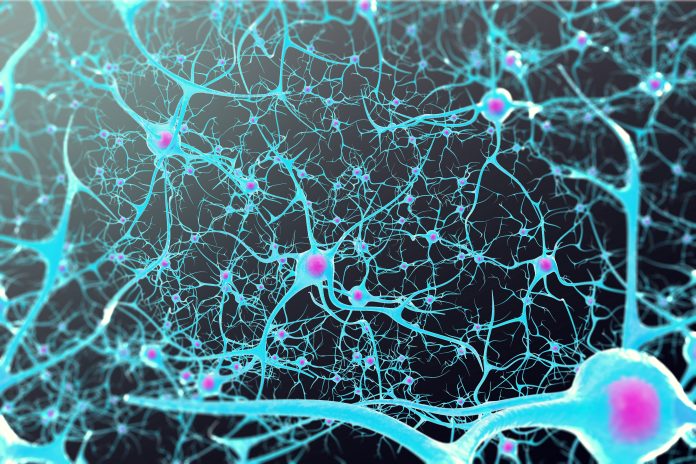
An analysis of the genetic profiles of thousands of neurons from postmortem brain tissue of people with amyotrophic lateral sclerosis (ALS) and from healthy donors has revealed how a set of genes could cause neuron loss in ALS and frontal temporal dementia (FTD). The study, which was funded by the National Institutes of Health (NIH) and led by researchers at the Broad Institute of MIT and Harvard, was published in Nature Aging.
Amyotrophic lateral sclerosis (ALS) is a neurodegenerative disorder characterized by a progressive loss of motor function linked to degenerating extratelencephalic neurons/Betz cells (ETNs). The result of this degeneration in the brain and spinal cord leads to muscle weaknesses paralysis, with most people diagnosed with ALS dying within three to five years.
The reasons why these neurons are selectively affected remain unclear. While previous research has identified a number of different genes that are associated with ALS most cases of the neurodegenerative disease are sporadic, have no known family history or etiology, which has made developing models of the disease a significant challenge.
But recent advances in single-cell analysis of brain tissue is beginning to yield new insights. In this study, “we applied single-nucleus RNA sequencing (snRNAseq) and in vitro human induced pluripotent stem cell modeling to investigate changes in cortical cell types in sporadic ALS (sALS),” the researchers wrote. This analysis found higher levels of risk ALS-FTD risk genes that were especially prominent in Betz cells, motor neurons that express the marker THY1. This was linked to disruptions in other neurons, which hindered their ability to build, transport, and break down proteins. The genes associated with this are SOD1, KIF5A, and CHCHD10, some of the most commonly identified genes associated with ALS and FTD.
“Our profiling identified the intrinsically higher expression of ALS–FTD risk factors in a specific class of extratelencephalic excitatory neurons,” the investigators noted. “In patients with ALS, these neurons and other subclasses of ETNs selectively express higher levels of genes connected to unfolded protein responses and RNA metabolism. We found that excitatory neuronal vulnerability is accompanied by a decrease in myelination-related transcripts in oligodendroglial cells and an upregulation of a reactive, proinflammatory state in microglia.”
Betz cell degeneration is a key indicator of ALS and is thought to occur early in ALS development when symptoms first appear. Developing a better understanding of neuron loss in ALS and why these and other cells become vulnerable in the disease has the potential to provide new therapeutic targets that could slow, or even halt, ALS progression.
In addition to ETNs, the investigators also sought to better understand how glial cells—cells that help support neuron health—are also affected by ALS. Prior research revealed that glial cells can become dysfunctional in ALS damaging neurons and hastening their death. For this study the team analyzed genetic data from two different kinds of glial cells and uncovered genes associated with cellular stress and inflammation. The researchers noted that the changes they observed also had an overlap with microglia in both multiple sclerosis and Alzheimer’s disease “suggesting that drugs modulating myeloid cells in neurodegenerative diseases may provide a basis for new therapeutic approaches.”
However, the team pointed out that more research is needed to determine if the glial cell dysfunction they observed is a consequence of neuron degeneration in ALS, or is one of the causes.
“Our study offers a view in which neurocentric disease vulnerability might spark responses in other cell types, but it also shows that enrichment of ALS–FTD-related genes in ETNs is coupled with processes engaging these genes in other cells too, that is, microglia,” the researchers concluded. “This view is a first insight into the disruptions of cortical biology in ALS and provides a connection between changes in cellular components and mechanisms associated with ALS. Future investigations should consider multicellular disruptions in ALS–FTD, where the survival of the neuron is unmistakably pivotal, but targeting other cells to reduce inflammation, promote myelination and bolster neuronal circuitry may re-establish a neuroprotective environment.”


